When studying immune cells, environment matters
Report urges researchers to look outside the petri dish and into the body to study T cell metaboli
October 10, 2019

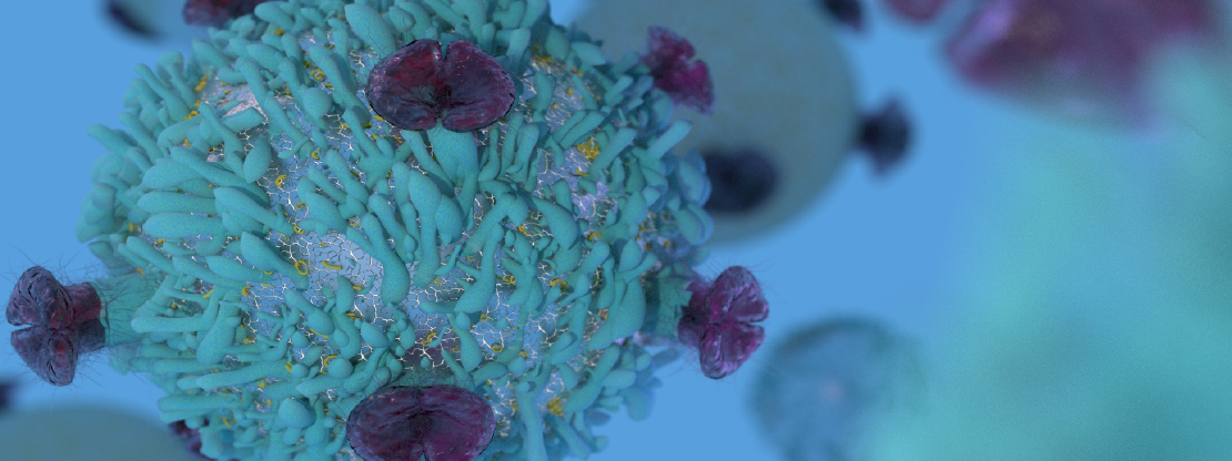
GRAND RAPIDS, Mich. (Oct. 10, 2019) — For years, scientists have used cells grown in petri dishes to study the metabolic processes that fuel the immune system. But a new report in Immunity suggests looking outside the dish and into living organisms gives a drastically different view of the way immune cells process and use energy.
Past research has shown that specialized immune cells called T cells convert a sugar called glucose into energy to power cellular function. However, this belief was largely built on data from cells grown in petri dishes, far removed from their normal environment.

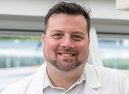
“It is akin to observing animal behavior in a zoo versus in the wild. Our immune cells don’t operate in a vacuum — they work in concert with a host of other cells and factors, which influence how and when energy is used,” said Russell Jones, Ph.D., the study’s senior author and head of Van Andel Institute’s Metabolic and Nutritional Programming group. “Understanding cellular metabolism is a critical piece of therapeutic development. Our findings reinforce the necessity of studying these cells in as close to a natural environment as possible.”
Jones and his colleagues found that T cells in a living system use glucose as building blocks for replicating DNA and other maintenance tasks, in addition to converting glucose into raw energy. They also discovered that the ways T cells process glucose evolves over the course of an immune response, which suggests T cells may use resources differently in the body when fighting a bacterial infection like Listeria or a disease like cancer.
The findings have far-reaching implications for how scientists study the complex, interconnected systems that underpin health and disease and how they translate these insights into new diagnostic and treatment strategies.
“Immune cells are far more dynamic in how they respond metabolically to infections and diseases than we previously realized,” Jones said. “For a while, we’ve been at a point in metabolism research that’s like standing in the dark under a street lamp — we could only see immediately in front of us. These findings will help us turn on the flood lights and illuminate the way to a more complete understanding of what immune cells need for optimal function.”
The findings were made possible thanks to a new method developed in consultation with collaborator Ralph DeBerardinis, M.D., Ph.D., that allowed Jones and his colleagues to map how T cells use nutrients under varying circumstances in living organisms.

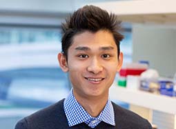
“Going forward, this new mapping technique will be invaluable as we pursue disease-specific studies,” said Eric Ma, Ph.D., the study’s first author and a postdoctoral fellow in Jones’ lab. “It has the potential to be transformative.”
In the future, the team plans to design human studies to measure how T cells use glucose and other nutrients when they are responding to pathogens or other insults like injury or diseases such as cancer.
Other authors include Kelsey S. Williams, Ph.D., Ryan D. Sheldon, Ph.D., and Connie M. Krawczyk, Ph.D., of Van Andel Institute; Mark J. Verway, Ph.D., Radia M. Johnson, Ph.D., Dominic G. Roy, Ph.D., Bozena Samborska, M.Sc., Takla Griss, Ph.D., Stephanie A. Condotta, Ph.D., and Martin J. Richter, Ph.D., of Goodman Cancer Research Centre, McGill University; Mya Steadman, Sebastian Hayes, Ph.D., Penelope A. Kosinski, Ph.D., Hyeryun Kim, Ph.D., Kelly Marsh, Ph.D., Victor Chubukov, Ph.D., and Thomas Roddy, Ph.D., of Agios Pharmaceuticals; and Brandon Faubert, Ph.D., and Ralph J. DeBerardinis, M.D., Ph.D., of Children’s Medical Center Research Institute, University of Texas Southwestern Medical Center. DeBerardinis also is affiliated with Howard Hughes Medical Institute. Van Andel Institute’s Flow Cytometry Core and Metabolomics Core and McGill University/Goodman Cancer Research Centre’s Metabolomics Core Facility also contributed to this work.
This research was supported by funding from Van Andel Institute (Krawczyk, Jones); McGill Integrated Cancer Research Training Program (Ma); Fonds de la Recherche du Québec–Sante (Ma, Roy, Jones); the Canadian Institutes of Health Research under grant no. MOP-142259 (Jones); and Agios Pharmaceuticals. The content is solely the responsibility of the authors and does not necessarily represent the official views of the funding organizations.
###
ABOUT VAN ANDEL INSTITUTE
Van Andel Institute (VAI) is an agile biomedical research and education organization that unleashes innovation to multiply impact. Established in Grand Rapids, Michigan, by the Van Andel family, VAI collaborates around the world to advance bold ideas in biomedicine and education. With a staff of more than 400 and a growing network of partners, VAI studies the origins of cancer, Parkinson’s and other diseases and translates its findings into breakthrough prevention and treatment strategies to improve human health. The Graduate School at VAI offers a Ph.D. in molecular and cellular biology through a research-intensive, interdisciplinary program that prepares students for successful careers as independent investigators. VAI also is dedicated to creating classrooms where curiosity, creativity and critical thinking thrive, offering engaging programs for K–12 students and transformative professional development and instructional tools for teachers. Find out more about Van Andel Institute by visiting vai.org.
Media Contact
Beth Hinshaw Hall
Van Andel Institute
[email protected]
616-234-5519
Media assets available: Investigator photo
