Your immune cells are what they eat
Salk Institute, Van Andel Institute scientists establish novel link between cell nutrition and identity, say targeting nutrient-dependent activity could improve immunotherapies NOTE: Originally published by Salk Institute
December 12, 2024

GRAND RAPIDS, Mich. (Dec. 12, 2024) — The decision between scrambled eggs or an apple for breakfast probably won’t make or break your day. However, for your cells, a decision between similar microscopic nutrients could determine their entire identity. If and how nutrient preference impacts cell identity has been a longstanding mystery for scientists — until a team led by Salk Institute and Van Andel Institute scientists revealed a novel framework for the complicated relationship between nutrition and cell identity.

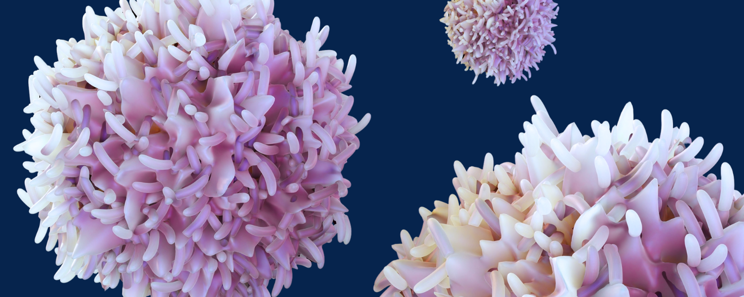
The answers came while the researchers were exploring different kinds of immune cells. The immune system relies on specialized “effector” T cells to fight off pathogens, but when fighting against cancer the perpetual activation of these T cells can make them “exhausted” and unable to continue fighting. In the new study, the team discovered that a nutritional switch from acetate to citrate plays a key role in determining T cell fates, shifting them from active effector cells to exhausted cells. This finding highlights how metabolic changes influence T cell identity and open avenues for interventions to sustain immune function against cancer.
The discovery that different nutrients can change a cell’s gene expression, function and identity significantly advances scientists’ understanding of the relationship between nutrition and cellular health throughout the body. It may also be possible to develop new therapies that target these nutrient-dependent mechanisms to help T cells stay active and energetically optimized against chronic diseases like cancer.
The findings were published in Science on Dec. 12, 2024.
“You know the saying, ‘You are what you eat?’ Well, we uncovered a way in which this actually operates in cells,” says Susan Kaech, Ph.D., senior author of the study and holder of the NOMIS Chair at Salk. “This is really exciting on two levels: on a fundamental level, our findings show that a cell’s function can be directly linked to its nutrition; on a more specific level, this sheds new light on how T cells become dysfunctional or exhausted and what we could do to prevent that.”

Metabolism is a central cellular process that transforms nutrients into metabolites and energy. Nutrients provide the resources for all cellular activities, but they must first be broken down into smaller molecules called metabolites. Metabolites have many uses, including promoting epigenetic regulation, a process that changes the shape of a cell’s DNA to alter the accessibility of different genes. Which genes are expressed in a cell at any given time then determines the behavior and identity of the entire cell.
The team wondered: Could this change in metabolism be responsible for the epigenetic changes that turn effector T cells into exhausted T cells? Is there a link between nutrition and exhausted T cell differentiation?
“Our findings reinforce the tightknit, nuanced relationship between nutrition, epigenetics and cell health,” said Russell Jones, Ph.D., a study author and chair of VAI’s Department of Metabolism and Nutritional Programming. “This work also paves the way toward improved immunotherapies against cancer and other chronic diseases.”
One of the most important and common metabolites is acetyl-CoA, which both effector and exhausted T cells make — but with one interesting difference. Exhausted T cells tend to make their acetyl-CoA using a protein called ACLY that uses citrate, rather than using a protein called ACSS2 that uses acetate.
The preferential activity of citrate-using-ACLY in exhausted T cells and acetate-using-ACSS2 in effector T cells piqued the team’s curiosity, leading them to genetically investigate the production of these metabolic proteins in both T cell subtypes. They found that ACSS2 gene expression was most highly expressed in functional T cells, but was drastically reduced in exhausted T cells in both mouse and human tissue samples. In contrast, ACLY genes were expressed similarly in both effector and exhausted T cells — with slightly greater expression in the exhausted cells. This suggested that T cells needed to express ACSS2 to maintain a functional state and that with exhaustion comes a greater reliance on ACLY.
To verify their findings, they went into the T cells and deleted ACLY and ACSS2 genes one at a time — discovering that the loss of ACLY boosted anti-tumor T cell activity, while the loss of ACSS2 did the opposite and reduced T cell efficacy. Seeing these differences in expression of ACLY and ACSS2 then led to a line of questioning about whether the downstream acetyl-CoA derived from these proteins may be determining the formation of exhausted T cells.
“We were shocked and thrilled to find the types of nutrients our cells were using changed their genetic expression and identities, meaning we have a whole new nutrient-dependent process to target with therapeutics that better equip us to fight chronic illness,” says Shixin Ma, Ph.D., first author of the study and postdoctoral researcher in Kaech’s lab.
Related: Learn more about research in the Russell Jones Lab ➔
Exhausted T cells were bailing on ACSS2 and relying heavily on ACLY, forcing themselves to use more citrate and less acetate to create acetyl-CoA, despite equal availability of both nutrients. Upon closer inspection, the researchers noticed that two distinct pools of otherwise identical acetyl-CoA molecules were piling up in different locations in the nucleus — where the cell’s DNA is stored — based on whether it was derived from acetate via ACSS2 or from citrate via ACLY. Each nutrient-specific pile was then linked to unique histone acetyltransferases, which are proteins that influence which genes are expressed to change cellular behavior and identity.
In a sort of domino effect, the original nutrient was ultimately determining T cell fate — (1) the metabolic enzyme (ACSS2 or ACLY) determined the nutrient used, (2) the metabolic enzyme determined the location of acetyl-CoA, (3) the location of acetyl-CoA determined what gene-modifying histone acetyltransferases were activated, and (4) those histone acetyltransferases either maintained the effector T cell identity or encouraged the shift to exhausted T cell identity.
This novel link between nutrition and cell identity offers a new explanation for exhausted T cell identity and in turn offers a multitude of new targets for future therapeutics that could keep T cells turned “on” longer.
“Truly, this is a radical concept that hasn’t been seen before,” says Kaech. “We are seeing clear consequences in cellular identity and function based on nutrient preferences by cells. The impact of these findings won’t just be within immunotherapy and immunology — every cell type in the body uses these metabolic processes, so plenty of other discoveries and therapeutic innovations can come out of what we’ve found.”
Today’s findings build on a related study by Jones and Kaech that described for the first time how immune cells leverage ACSS2 and ACLY to produce acetyl-CoA.
Other authors include Thomas Mann, Steven Zhao, Bryan McDonald, Yagmur Farsakoglu, Lizmarie Garcia-Rivera, Filipe Araujo Hoffmann, Shihao Xu, Victor Du, Dan Chen, Jesse Furgiuele, Michael LaPorta, and Emily Jacobs of Salk; Michael Dahabieh, Lisa DeCamp, Brandon Oswald, Ryan Sheldon, and Abigail Ellis of Van Andel Institute; Won-Suk Song and Cholsoon Jang of UC Irvine; H. Kay Chung of Salk and the University of North Carolina at Chapel Hill; Longwei Liu and Yingxiao Wang of the University of Southern California; and Peixiang He of UC San Diego.
Research reported in this publication was supported by the National Institute of Allergy and Infectious Diseases of the National Institutes of Health under award nos. R01AI066232 (Kaech), R21AI151986 (Kaech) and R01AI165722 (Jones); National Institute on Alcohol Abuse and Alcoholism of the National Institutes of Health under award no. R01AA029124 (Jang); National Institute of Biomedical Imaging and Bioengineering of the National Institutes of Health under award no. R01EB029122 (Wang); National Institute of General Medical Sciences of the National Institutes of Health under award no. R35GM140929 (Wang); the National Cancer Institute of the National Institutes of Health under award nos. K00CA222741 (Zhao), P30CA01495 (Salk Institute Flow Cytometry Core Facility), and T32CA009370 (Mann); the National Institutes of Health Office of the Director under award nos. S10-OD023689 (Salk Institute Flow Cytometry Core Facility) and S10OD034268 (Salk Institute Flow Cytometry Core Facility); the National Institute on Aging of the National Institutes of Health under award no. P30AG068635 (Gage); Chapman Foundation and the Helmsley Charitable Trust; an Allen Distinguished Investigator Award, a Paul G. Allen Frontiers Group advised grant of the Paul G. Allen Family Foundation (Jones); Van Andel Institute (Jones); American Association for the Study of Liver Diseases Foundation (Jang); Edward Mallinckrodt Jr. Foundation (Jang); Cancer Research Institute (Mann and Ma); Damon Runyon Cancer Research Foundation (Mann); and a Salk Pioneer Fund Postdoctoral Scholar Award (Ma). The content is solely the responsibility of the authors and does not necessarily represent the official views of the National Institutes of Health or other funders.
